


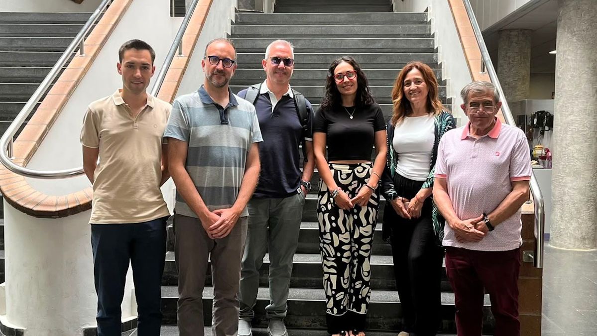
A team from the Universitat Politècnica de València (UPV) and the University of Valencia (UV) has developed and validated an artificial intelligence (AI) system capable of distinguishing Ewing sarcoma from other morphologically similar tumours with high accuracy. The model, which also enables the full optimisation of tissue use in biopsies, achieves 91.6% accuracy.
The breakthrough, published in the journal International Journal of Molecular Sciences, represents a leap forward in digital pathology by aiding the diagnosis of complex paediatric and juvenile neoplasms; it reduces errors and preserves the scarce biological material obtained during needle biopsies.
Ewing sarcoma is a rare malignant tumour that primarily affects children, adolescents and young adults. It usually occurs in the bone, although it can also develop in soft tissues. One of the main difficulties in diagnosing it is that, under the microscope, it can be very similar to other tumours that also originate in soft tissues, such as rhabdomyosarcoma (striated skeletal muscle), chondrosarcoma (cartilage), GIST/gastrointestinal stromal tumours (the digestive tract), or synovial sarcoma (joints).
Researchers from the Department of Pathology at the University of Valencia – Francisco Giner (lead researcher and first author of the article), Isidro Machado and Samuel Navarro (also coordinator of the paediatric solid tumour translational research group at the INCLIVA Institute for Health Research), said, “in many small biopsies, there is very little tissue. If the pathologist has an initial tool that accurately guides the suspected diagnosis, they can decide which additional and immunohistochemical tests to request, thereby avoiding using up the sample on analyses that do not provide decisive information.”
The development of the new AI tool was led by a team from the CVBLab research group at the Universitat Politècnica de València (UPV) and the company Artikode Intelligence, a spin-off from the UPV.
“The tool is designed to support medical staff in the field of anatomical pathology and could help guide the tests needed to confirm the diagnosis, particularly when very little tissue is available from a biopsy. Using deep learning techniques, the system identifies the microscopic patterns associated with each type of tumour and subsequently generates a classification,” said Valery Naranjo, coordinator of the CVBLab at the UPV and CTO of Artikode Intelligence.
To train and evaluate the system, the researchers used 1,926 digitised histological samples from 729 patients at four hospitals in Spain and Italy. These included 517 cases of Ewing sarcoma, as well as other tumours.
The system achieved an overall diagnostic accuracy of 91.6% and a sensitivity of 97.1% specifically for Ewing sarcoma. The model had a marginal error rate of 1.96% between Ewing sarcoma and rhabdomyosarcoma, providing a reliable solution to the most challenging differential diagnosis among small round cell tumours.
Giner said: “The novelty of the project does not lie simply in using AI to diagnose Ewing sarcoma, as experimental deep learning approaches already exist in this field. Our distinctive contribution lies in the combination of the clinical problem addressed, the type of data, the classification strategy and the focus on developing a tool applicable to real-world digital pathology.”
He said AI is not intended to replace the pathologist, but rather to provide objective diagnostic support, which is particularly valuable in hospitals that do not have immediate access to the full range of molecular tests.